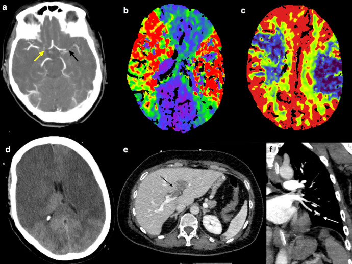
Fig. 3. Radiological findings in Patient 2.
a Computed tomograghy angiography (CTA) showed the proximal M1 segment occlusion of the left middle cerebral artery (MCA) (black arrow) and the occlusion of the right internal carotid artery terminus (yellow arrow); b CT perfusion (CTP) showed bilateral infarct core (cerebral blood volume—CBV—map in c) and hypoperfusion (mean transit time—MTT—map in b), without treatable penumbra; d 24 h after, brain CT revealed bilateral malignant MCA infarcts; e portal vein thrombosis with extension to the left intrahepatic branches (black arrow); f subsegmental pulmonary artery thrombosis (white arrow).

