. 2021 Jun 25;38(2):205–218. doi: 10.5511/plantbiotechnology.21.0222a

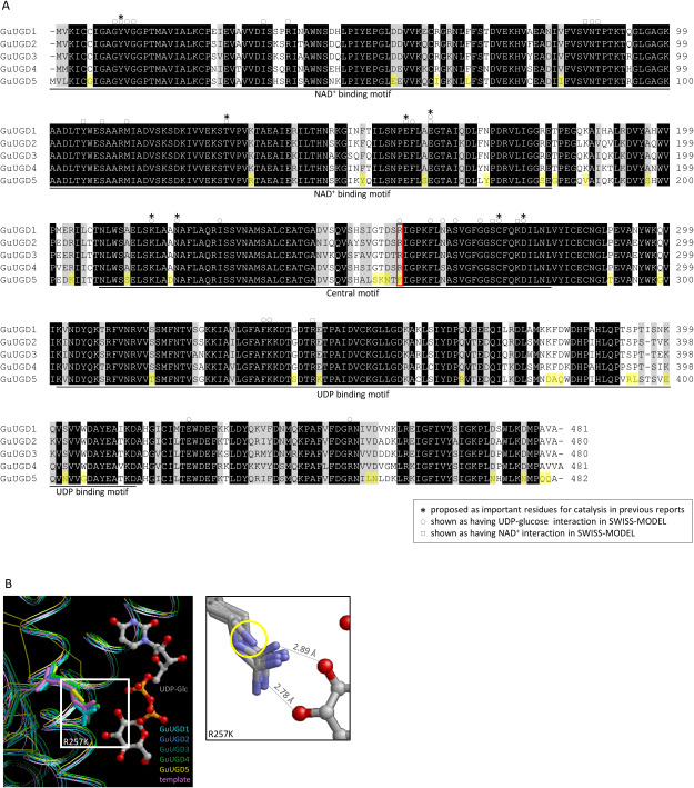
Figure 2. Amino acid sequences and predicted molecular structures of GuUGDs. (A) Alignment of the amino acid sequences deduced from cDNA of cloned GuUGDs. We made the alignment using BioEdit with ClustalW. The NAD+ binding motif, UDP binding motif, and central motif were annotated based on GenBank. Asterisks indicate key residues for catalysis proposed in previous studies (Campbell et al. 2000; Egger et al. 2011); circles and squares indicate amino acids shown as key residues interacting with UDP-glucose and NAD+, respectively, by SWISS-MODEL. Amino acid residues shown in yellow background were specific to GuUGD5. The amino acid residue framed in red was near UDP-glucose in the 3D protein models. (B) The 3D protein model of GuUGDs built with human UGD homo-hexamer structure as a template in SWISS-MODEL. Squares indicate the residue position that likely prevented UGD activity in GuUGD5; this position is indicated by a yellow circle in the enlarged panel (right). Atoms of oxygen, nitrogen, carbon, and phosphorous are indicated in red, blue, gray, and orange, respectively. Distances between substrates and the crystal structure of human UGD are displayed in the enlarged panel.

Figure 2. Amino acid sequences and predicted molecular structures of GuUGDs. (A) Alignment of the amino acid sequences deduced from cDNA of cloned GuUGDs. We made the alignment using BioEdit with ClustalW. The NAD+ binding motif, UDP binding motif, and central motif were annotated based on GenBank. Asterisks indicate key residues for catalysis proposed in previous studies (Campbell et al. 2000; Egger et al. 2011); circles and squares indicate amino acids shown as key residues interacting with UDP-glucose and NAD+, respectively, by SWISS-MODEL. Amino acid residues shown in yellow background were specific to GuUGD5. The amino acid residue framed in red was near UDP-glucose in the 3D protein models. (B) The 3D protein model of GuUGDs built with human UGD homo-hexamer structure as a template in SWISS-MODEL. Squares indicate the residue position that likely prevented UGD activity in GuUGD5; this position is indicated by a yellow circle in the enlarged panel (right). Atoms of oxygen, nitrogen, carbon, and phosphorous are indicated in red, blue, gray, and orange, respectively. Distances between substrates and the crystal structure of human UGD are displayed in the enlarged panel.