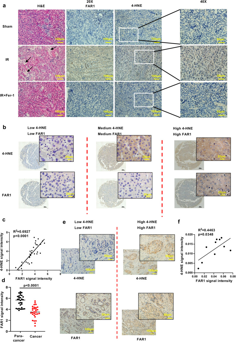
Fig. 5. FAR1 responds to the process of ferroptosis in renal IR and tumors.
a Representative immunohistochemical staining (IHC) of cortical renal tissues from the mice after ischemia reperfusion (IR) or sham treatment. The experiment was repeated three times, independently, with similar results. b Representative immunohistochemical staining (IHC) of liver cancer samples. The experiment was repeated twice, independently, with similar results. c Dot plot depicting the relevance of the signal intensity of FAR1 and 4-HNE IHC staining in the indicated liver cancer samples. d FAR1 signaling intensity in para-cancer and liver cancer tiusses indicates FAR1 is lower expressed in liver cancer tissues. e Representative immunohistochemical staining (IHC) of FAR1 and 4-HNE in human breast cancer samples. The experiment was repeated twice, independently, with similar results. f Dot plot depicting the relevance of the signal intensity of FAR1 and 4-HNE IHC staining in the breast cancer samples.

