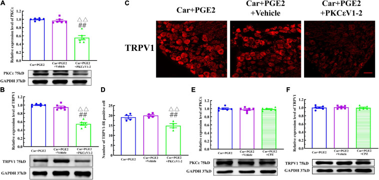
FIGURE 4.
Protein kinase C epsilon (PKCε) regulates TRPV1 expression in the DRG. (A) The quantification of the Western blot results and a representative Western blot showing inhibited PKCε protein isolated from the DRG 48 h after PGE2 injection. (B) The quantification of the Western blot results and a representative Western blot showing decreased TRPV1 protein isolated from the DRG 48 h after PGE2 injection. (C) TRPV1 staining in the peripheral nervous system 48 h after PGE2 injection. Scale bar 100 μm. (D) The quantification of TRPV1–IR positive neurons. (E) The quantification of the Western blot results and a representative Western blot showing PKCε protein isolated from the DRG 4 h after PGE2 injection. (F) The quantification of the Western blot results and a representative Western blot showing TRPV1 protein isolated from DRG 48 h after PGE2 injection. △△ Compared with the NS+PGE2 group, P < 0.01; ## compared with the Car+PGE2 group, P < 0.01.

