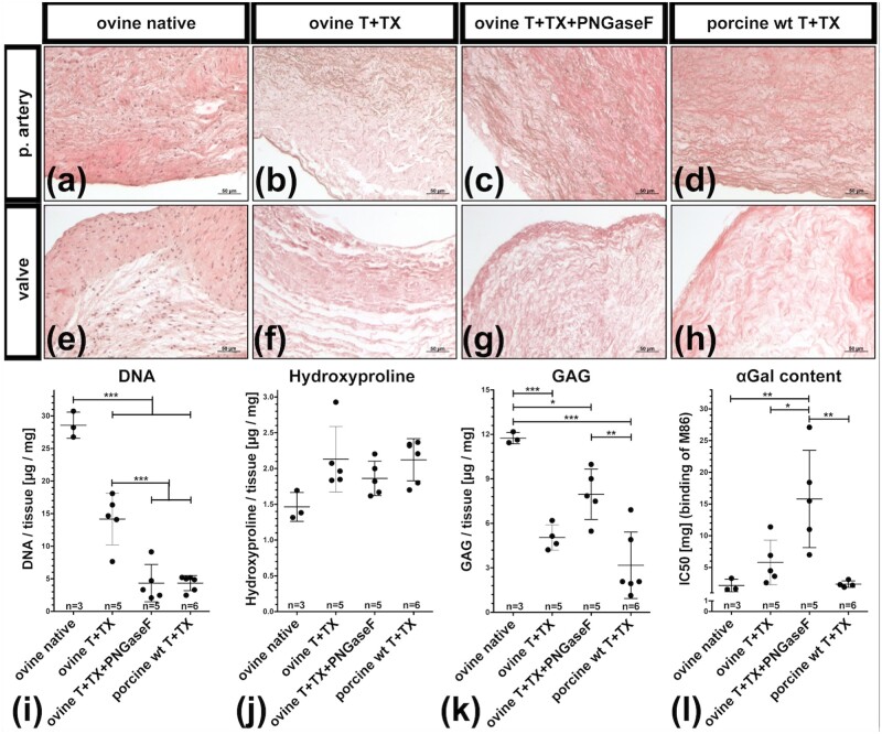
Figure 3.
Histological and biochemical characterization of decellularized valves (a–h) H&E-stained sections showing pulmonary arteries (p. artery) and valve cusps (valve) of native ovine PHVs (a, e), trypsin+TX decellularized ovine PHVs before (b–f) and after (c–g) PNGaseF treatment and trypsin+TX decellularized porcine PHVs (d, h). DNA (i), hydroxyproline (j) and GAG (k) contents were quantified in freeze-dried tissues. (l) The αGal epitopes in fresh tissues are depicted as IC50, determined an inhibitory ELISA. Low IC50 corresponds to high amounts of αGal, whereas high IC50 corresponds to low amounts of αGal epitopes present in the tested tissue. (i–l) Each dot represents an individual biological replicate. Scale bars represent 50 µm.

