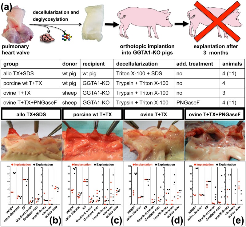
Figure 4.
(a) Schematic depiction of the experimental set-up, gross morphology and TEE results of dPHVs, orthotopically implanted into pigs for 3 months. TEE analysis was performed shortly after implantation and immediately prior to death. Valve diameter [mm], ejection fraction (EF) [%], transvalvular gradients (mean and max) [mmHG], grade of insufficiency and stenosis [0–4, 4 = total] and orifice area [cm2] were determined. (b) Porcine pulmonary grafts derived from wt pigs and decellularized with the aid of TX+SDS after 3 months implanted into wt pigs (allo) TX+SDS). (c) porcine pulmonary heart valves obtained from wt pigs and decellularized with the aid of T+TX 3 months after implantation into GGTA1-KO pigs (porcine wt T+TX). (d) xenogeneic ovine PHVs decellularized with T+TX after 3 months implanted into GGTA1-KO pigs (ovine T+TX). (e) xenogeneic ovine PHVs decellularized with T+TX and additionally treated with PNGaseF 3 months after implantation into GGTA1-KO pigs (ovine T+TX+PNGaseF).

