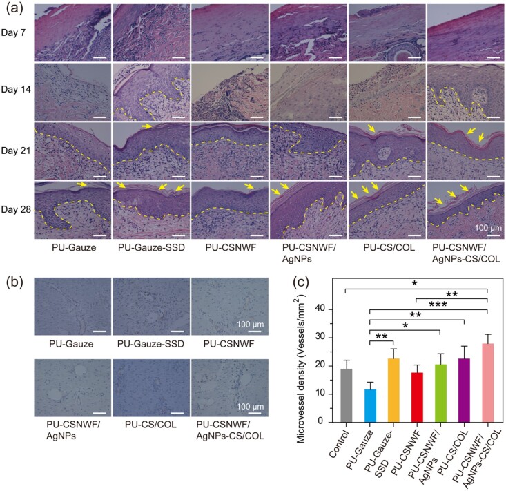
Figure 7.
Reepithelialization and angiogenesis in wound sites. (a) Representative images of histological sections with H&E staining on Day 7, Day 14, Day 21 and Day 28 post-surgery. The yellow dashed line indicates the boundary between the newly formed epidermis and dermis. The yellow arrow represents the stratum corneum. (b) Representative images of immunohistochemical staining of wound sections at Day 28 with factor VIII. (c) Quantification of vessel number per microscopic field in different groups (*P < 0.05, **P < 0.01 and ***P < 0.001)

