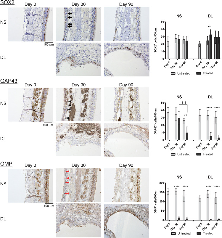
FIGURE 3.
Representative images of sections of the nasal mucosa stained with antibodies against OMP, SOX2, and GAP43 (magnification, 200×). OMP+ mature olfactory receptor neurons (ORNs), SOX2+ ORN progenitors, and GAP43+ immature ORNs in the epithelium were identified by immunohistochemical staining (brown). On day 30, mature OMP+ ORNs were sparse (red arrowheads), whereas GAP43+ immature ORNs (black arrowheads) were more frequently observed in the epithelium of the nasal septum (NS). In the upper lateral (DL) area, SOX2+ cells (black arrows) but no GAP43+ or OMP+ cells were present in the epithelium. On day 90, neither GAP43+ immature ORNs nor OMP+ mature ORNs were present in the epithelium of the NS or DL area. Graphs show the number of each cell type per 500 μm of basal layer length. Asterisks indicate a significant difference between the treated and untreated groups. Daggers indicate a significant difference between time points. **P < 0.01; ****P < 0.0001, ††††P < 0.0001 (n = 6, two-way analysis of variance).

