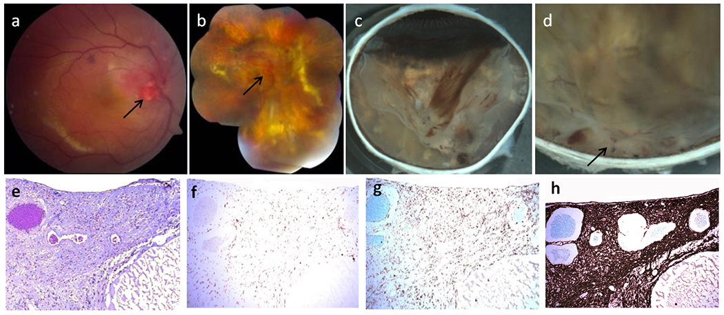
Fig. 1.

Clinical photos and photomicrographs in Patient 1 with von Hippel-Lindau (VHL)-associated retinal hemangioblastoma (RH). Fundus photographs of VHL-associated RH in the right eye taken in January 2005 (a) and March 2010 (b). a The baseline fundus photograph before the intravitreal ranibizumab trial showed a prominent optic nerve tumor (arrow), focal hemorrhages and yellowish retinal hard exudates. b Ranibizumab and radiation therapy shows multiple vitreous hemorrhages, enlarged optic nerve tumor, serous retinal detachment, lipid accumulation and multiple pigmentary/yellowish chorioretinal lesions in the retina. Macroscopic photographs of VHL-associated RH in the right eye after enucleation in January 2012 (c, d). c The inferior calotte of the globe showed several patches of vitreous hemorrhages, yellowish chorioretinal lesions and retinal detachment. d. A whitish mass with focal hemorrhages around the optic nerve head (juxtapapillary RH) and several smaller lesions are observed in the peripapillary retina. e A large juxtapapillary RH was noted at the optic nerve head with a huge cyst (C) (hematoxylin and eosin (H&E) stain, original magnification, ×100). Avidin-biotin-complex immunohistochemistry staining corresponding to the same section with panel shows CD68 positive macrophages (f) and CD45RO positive T lymphocyte (g) infiltration in the juxtapapillary RH (original magnification, ×100). h Marked gliosis (glial fibrillary acidic protein positive) occurs in the whole areas of hemangioblastoma and retina (Avidin-biotin-complex immunohistochemistry, original magnification, ×200)
