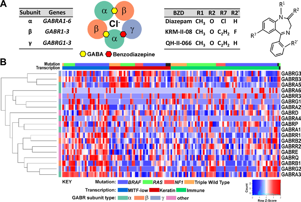
Figure 1. GABAA receptor expression in metastatic melanoma.
(A) Type-A GABA receptors (GABAARs) are composed most commonly of two α, two β, and γ subunits encoded by GABR genes GABRA (1–6), GABRB (1–3), and GABRG (1-3), respectively. GABAAR consists of five subunit transmembrane segments which create the chloride (Cl−) conduction pore. Inter-subunit binding sites for GABA (yellow hexagon) and benzodiazepine (red hexagon) are shown, recognizing the αβαβɣ subunit stoichiometry. Benzodiazepines have a common core structure. Shown are sites frequently modified (R1, R2, R2′, R7), which may impart a GABAAR subtype-preference. GABAAR subtype-preferring benzodiazepines (BZDs) KRM-II-08 and QH-II-066 differ from diazepam by having an R7 acetylene group. (B) Normalized expression data for GABR genes from stage III/IV melanoma specimens. Samples were classified into three melanoma molecular subgroups. Heatmap for analysis of expression across subgroups was generated using Morpheus (https://software.broadinstitute.org/morpheus).

