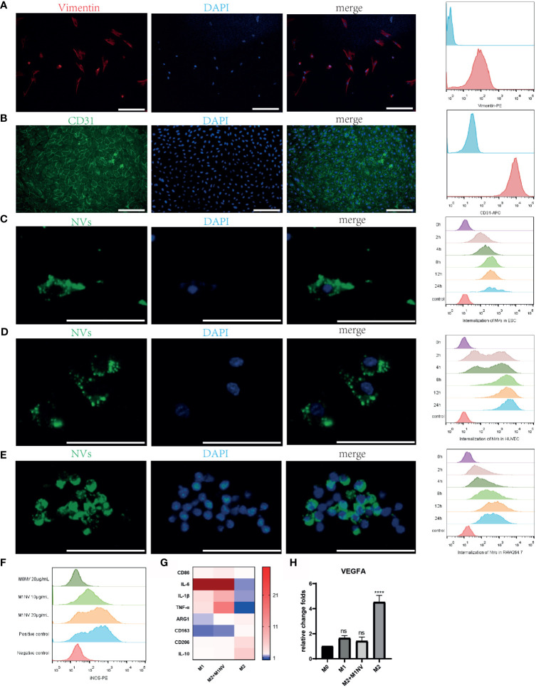
Figure 2.
M1NVs were internalized by primary cells and macrophages and reprogrammed M2 into M1 macrophages. (A) ESCs derived from endometrium expressed stroma marker Vimentin (Scale bar = 100 μm). (B) HUVECs derived from normal human umbilical veins expressed CD31, the endothelial marker (Scale bar = 100 μm). (C–E) NVs labeled by PKH67 were internalized in the ESCs, HUVECs and macrophages. With the passage of time, the number of NVs internalized into the cells was increasing (Scale bar = 100 μm). (F) M2 macrophages were polarized into M1 by M1NVs, and 20μg/mL of M1NVs were sufficient to play the role. (G) RT-qPCR revealed the up-regulated M1 markers (CD86, IL6, IL1B, and TNFA) and down-regulated M2 markers (ARG1, CD163, CD206, IL10) in Re-M2 macrophages. (H) Re-M2 macrophages expressed lower level VEGFA than M2 macrophages. ns, no significance when compared to M0. ****P<0.0001 when compared to M0.

