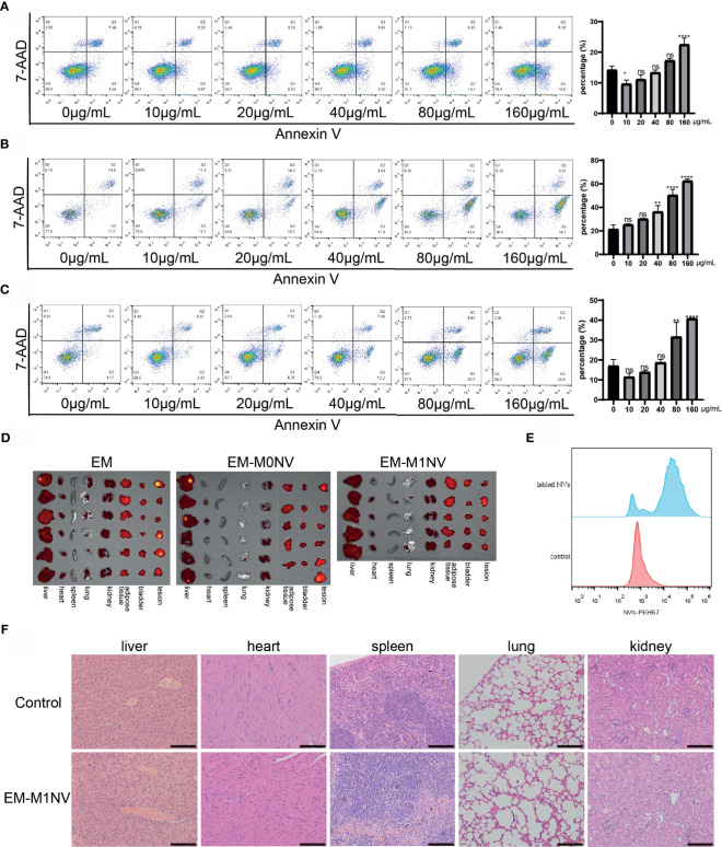
Figure 7.
M1NVs treatment did not induce cell apoptosis and did not injury the organs. (A) The apoptosis percentage of macrophages treated with different concentration of M1NVs. (B) The apoptosis percentage of ESCs treated with different concentration of M1NVs. (C) The apoptosis percentage of HUVECs treated with different concentration of M1NVs. ns, No significance when compared with the group treated with 0 μg/mL M1NVs. *P<0.05, **P<0.01, ****P<0.0001 when compared with group treated with 0 μg/mL M1NVs. (D) IVIS of the organs from different groups. Group EM was the negative control since no labeled NVs were injected. (E) FCM showed NVs labeled with PKH67 were engulfed by the peritoneal macrophages. (F) H&E staining sections of organs from control group and EM-M1NV group (Scale bar = 100 μm).

