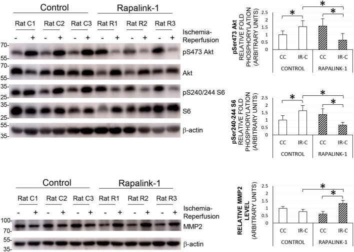
Figure 4.
The representative Western blots of pS6, pAkt, and MMP2, and their quantification after 1 h of MCAO and 2 h of reperfusion. ANOVA followed by multiple comparisons with the Bonferroni correction was used for the quantification of protein. The Phosphorylation of Akt at Ser473 and the phosphorylation of S6 at Ser240/244 were increased during ischemia–reperfusion that indicates the increased activity of mTORC2 and mTORC1, respectively. The treatment with Rapalink-1 resulted in a more than 50% decrease in the phosphorylation of both Akt and S6 in the IR-C when compared with the control rats, which suggests that Rapalink-1 inhibited both mTORC1 and mTORC2 in this experiment. The increased protein level of MMP2 with Rapalink-1 treatment in the IR-C suggests that Rapalink-1 could aggravate blood–brain barrier (BBB) disruption in the ischemic-reperfused area. CC, contralateral cortex; IR-C, ischemic-reperfused cortex; Control, MCAO/reperfusion group; Rapalink-1, MCAO/reperfusion + Rapalink-1 group. n = 6–9. *p < 0.05. Values are means ± SD.

