
Fig. 4.
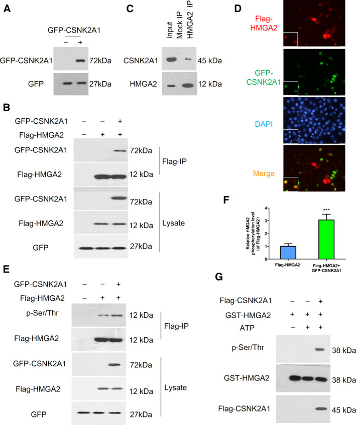
The relationship between HMGA2 and CSNK2A1. (A) Confirmation of the plasmid of GFP‐CSNK2A1 by a western blot assay. (B) 293T cells co‐transfected with indicated plasmids were lysed and immunoprecipitated using Flag‐M2 beads followed by a western blot assay with anti‐GFP and anti‐Flag antibodies. (C) An endogenous HMGA2‐CSNK2A2 complex was verified in HeLa cells. HeLa cells extracts were immunoprecipitated against HMGA2 or IgG isotype antibodies (Mock IP) and were analyzed by immunoblotting using anti‐CSNK2A1 antibody. (D) HeLa cells co‐transfected with Flag‐HMGA2 and GFP‐CSNK2A1 were stained for Flag (red) and GFP (green), visualized by fluorescence microscopy, and nuclei were stained with DAPI. (E) 293T cells transfected with indicated plasmids were lysed and immunoprecipitated using Flag‐M2 beads and a western blot assay was used for phosphorylation of HMGA2 by anti‐p‐Ser/Thr antibody. (F) Relative HMGA2 phosphorylation levels were quantified using imagej. (G) An in vitro phosphorylation assay showed that CSNK2A1 phosphorylated HMGA2. Data were analysed using Student's t‐test and are presented as the mean ± SD of three independent experiments. ***P < 0.001.