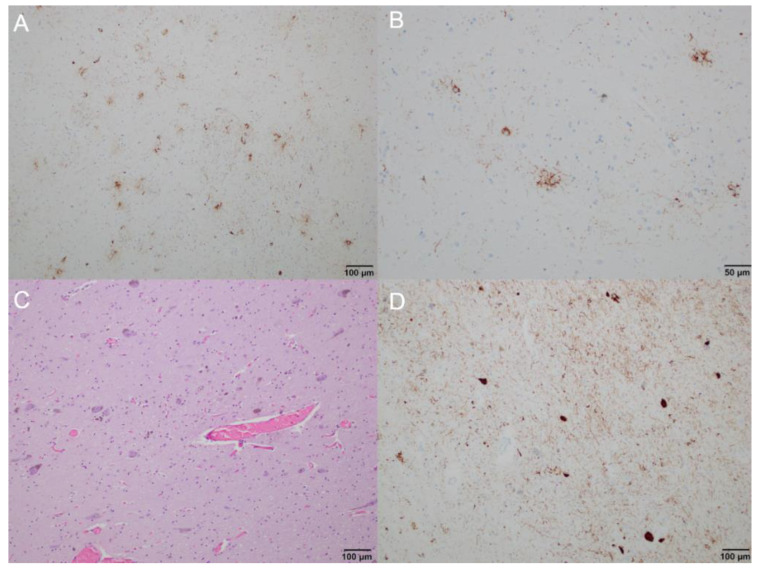
Figure 2.
Brain neuropathology, microscopic anatomy: (A) immunohistochemistry staining showing tau accumulation in the putamen, predominantly in the form of tufted astrocytes and tau-positive neurons (100× magnification); (B) immunohistochemistry staining showing tufted astrocytes in the caudate nucleus (200× magnification); (C,D) substantia nigra showing neuronal cell loss, tau-positive neurons, and numerous neuropil threads ((C) hematoxylin and eosin staining; (D) immunohistochemistry staining; 100× magnification).

