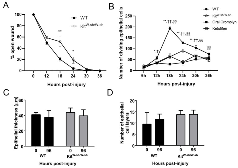
Figure 5.
Changes in epithelial healing after corneal abrasion in KitW-sh/W-sh mice and WT mice treated with mast cell stabilizers. (A) The rate of epithelial wound closure was determined by fluorescein dye retention and expressed as a percent of the initial wound area. (n = 6 per group, * p ≤ 0.05, ** p ≤ 0.01). (B) Numbers of dividing basal epithelial cells were determined at different times after corneal abrasion. (n = 6 per group, * p ≤ 0.05, ** p ≤ 0.01 compared to KitW-sh/W-sh; n = 6 per group, † p ≤ 0.05 and †† p ≤ 0.01 compared to oral cromolyn; n = 6 per group, §§ p ≤ 0.01 compared to ketotifen). (C) Epithelial thickness and (D) numbers of epithelial cell layers were determined from transverse histological sections of the cornea before injury (time 0) and 96 h post-abrasion.

