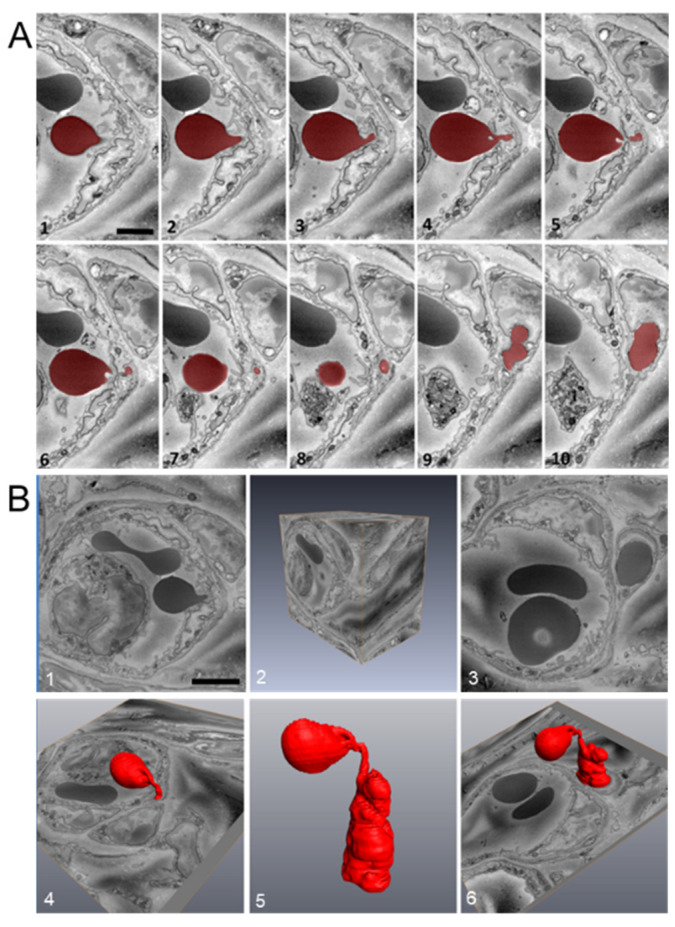
Figure 8.
RBC extravasation after corneal abrasion. (A) Sequential images (1–10) were acquired using serial block-face scanning electron microscopy (SBF-SEM) and used to confirm RBC (colored red) passage across the venular endothelium. (B) Three-dimensional reconstruction analysis of the complete image stack (part of which is shown in panel (A)) illustrates the high degree of RBC deformability (panel B5) needed to pass through the endothelium.

