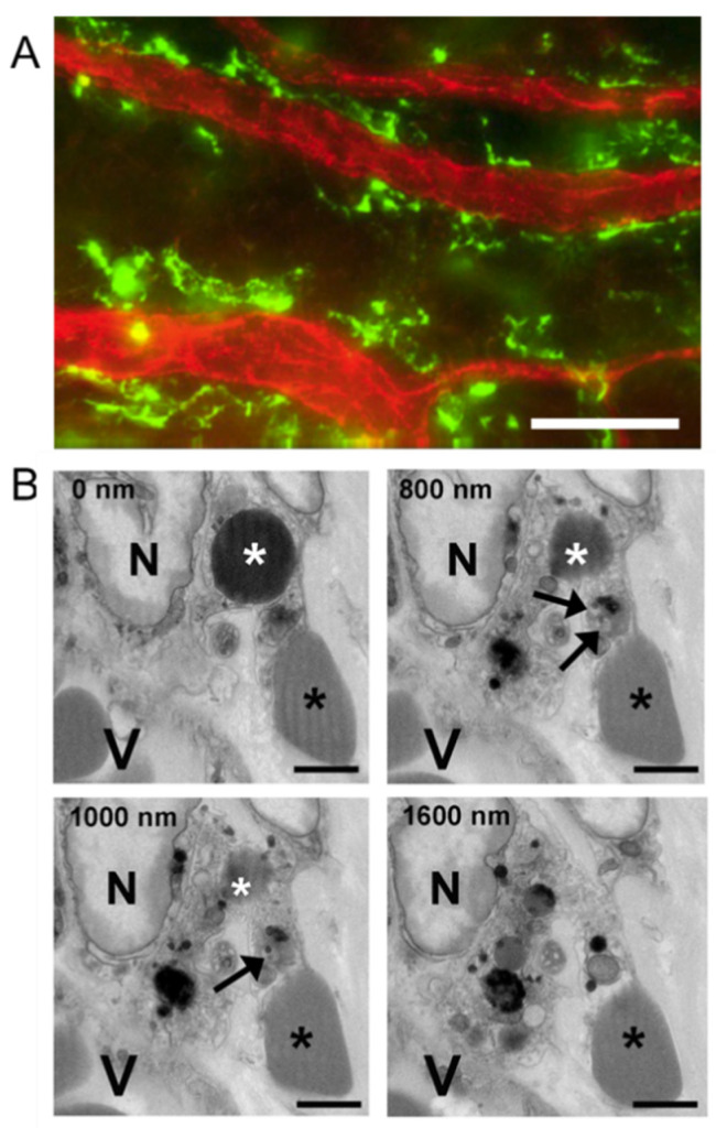
Figure 9.
Perivascular macrophages and RBC and platelet phagocytosis after corneal abrasion. (A) Immunofluorescence image of perivascular macrophages (green, anti-CD301) lying next to limbal venules (red, anti-CD31). (B) Sequential SBF-SEM images reveal a perivascular macrophage containing a phagocytosed RBC and platelet. Top left in each panel indicates the depth of sectioning in nanometers. In each panel, a large macrophage nucleus (N) can be seen and the macrophage is located near a blood vessel (V). A phagocytosed RBC (white asterisk) is visible at 0, 800, and 1000 nm depths within the macrophage cytoplasm, but not at 1600 nm. A phagocytosed platelet containing dense granules (black arrows) is visible at 800 and 1000 nm depths but is absent at 0 and 1600 nm. An extravascular RBC (black asterisk) is in contact with the macrophage throughout the series. Bar = 25 µm (A); Bar = 2µm (B).

