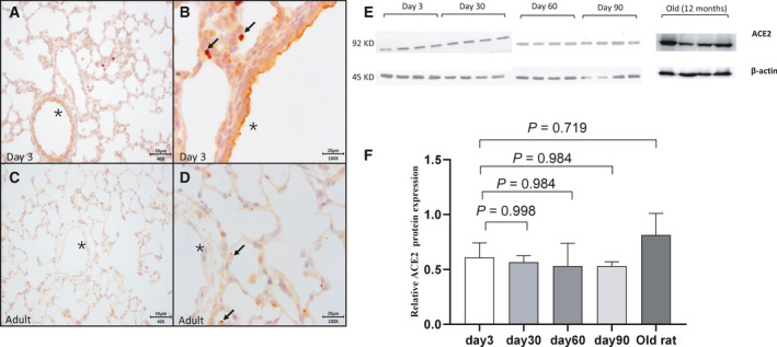
Fig. 1.

Immunohistochemistry staining and pulmonary protein level of ACE2 in rat’s lung tissues at different ages (Fig. 1A,B for neonatal lungs, Fig. 1C,D for adult lungs). In neonates, cells stained positively for ACE2 were few in neonatal lungs (Fig. 1B, arrows), while ACE2 was stained extensively in the adult lung (Fig. 1C) with a special profile of polarization to the alveolar space (Fig. 1D, arrows). Pulmonary endothelium was stained positive for ACE2 in neonatal rather than adult lung (Fig. 1B,D, stars). Scale bars, 50 μm in panel A and C; 20 μm in panel B and D. A total amount of 30 μg protein was loaded to the gel, and β‐actin was used as a reference (Fig. 1E,F N = 4). Each group was compared with the group of Day 3 using one‐way ANOVA, followed by Dunnett multiple comparisons test. (Fig. 1F). The bars were presented as mean with standard error of the mean (SEM).
