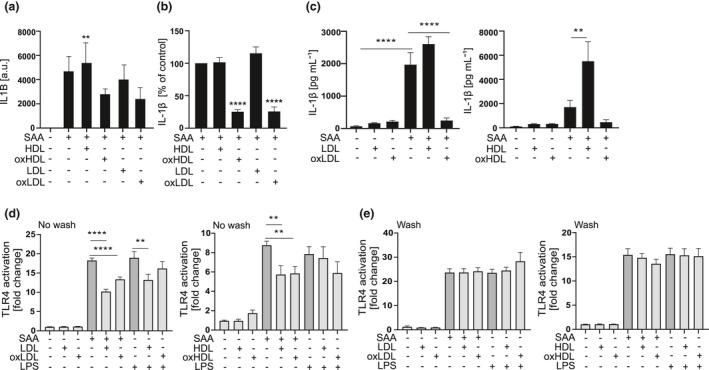
Figure 3.

Inhibition by oxidised lipoproteins does not require binding to SAA. (a) Primary macrophages were subjected to native or oxidised HDL3 and LDL (400 μg mL−1) for 1 h, washed and changed into fresh SFMM containing SAA (3 μg mL−1), and incubation was continued for 5 h. IL1B mRNA levels were analysed by quantitative real‐time RT‐PCR relative to GAPDH. Data are expressed as arbitrary units (a.u.) relative to GAPDH. (b) Primary macrophages were subjected to native or oxidised HDL3 and LDL (400 μg mL−1) for 1 h, washed and changed into fresh SFMM containing SAA (3 μg mL−1) and incubated for 18 h. Cell culture media were analysed for IL‐1β concentrations by ELISA. (c) THP‐1 macrophages were subjected to native or oxidised HDL3 and LDL (200 μg mL−1) for 1 h and then to SAA (3 μg mL−1) for 5 h. Secretion of IL‐1β was analysed from cell culture media by ELISA. HEK‐blue TLR4 reporter cells were changed to SFMM before addition of native or oxidised HDL3 and LDL (lipoproteins), (d) no wash group, or after 1‐h lipoprotein activation, before SAA was applied, (e) wash group, to remove the lipoproteins. SEAP activity was detected from supernatants 24 h after addition of lipoproteins. The data are presented as fold changes compared to separate untreated cells. Data are means from 2 (a) or at least 3 (b) or 4 (c–e) independent experiments. **P < 0.01, ****P < 0.0001.
