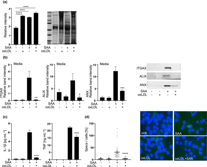
Figure 6.

Oxidised LDL inhibits the NLRP3 inflammasome‐induced vesicle secretion and ASC oligomerisation. (a) THP‐1 macrophages were treated with oxLDL 200 µg mL−1 for 1 h and challenged with SAA (3 μg mL−1) for 5 h. Silver‐stained SDS‐PAGE gel was used to estimate the total protein secretion. On the left average of the relative lane volumes are shown, on the right representative silver staining is shown. (b) Secretion of ITGAX, alix and annexin‐1 (ANX) was analysed by Western blotting. Equal volumes of media were loaded, on the right representative blots are shown. (c) Secretion of IL‐1β and TNF was analysed from media samples used for analysis of extracellular vesicle secretion and are expressed as ng mL−1 of IL‐1β (left panel) or TNF (right panel). (d) THP‐1 macrophages were pretreated with oxLDL 400 µg mL−1 for 1 h and activated with SAA (3 μg mL−1) for 5 h and ASC oligomerisation was visualised by stainings, ASC (green) and nuclei (blue, DAPI). On the left: speck formation was quantified by counting the number of specks per field, which was divided by the number of nuclei per field. On the right: stainings are representatives of four independent experiments. Mean values shown are from 4 (a–d) independent experiments. *P < 0.05, **P < 0.01, ***P < 0.001, ****P < 0.0001.
