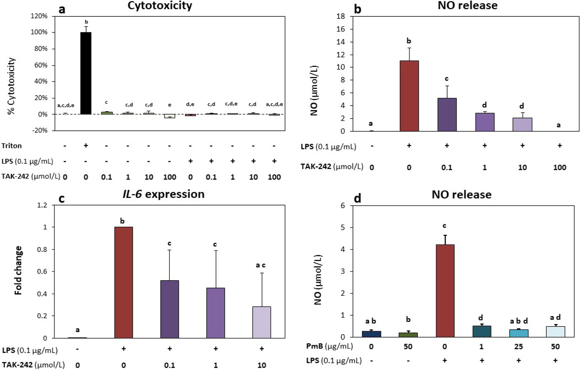
Fig. 2. TLR4 inhibition mitigated LPS-induced inflammatory response.

(a) Cytotoxicity (%) based on LDH levels did not increase with either LPS (0.1 μg/mL) or TAK-242 treatment, indicating no loss in cell viability in any of the experimental treatment group. Cells treated with 10× Triton X-100 for 10 min at the end of culture were used as a cytotoxic (kill/positive) control. (b) TAK-242 inhibited LPS-induced increases in NO release in a dose-dependent manner. (c) Cells treated with LPS showed significantly up-regulated expression of IL-6; TAK-242 co-treatment significantly decreased IL-6 expression when compared to LPS-only group. Gene expression data shown as fold change versus LPS-only group. (d) NO release increased in cells treated with LPS. PMB inhibited LPS-induced increase in NO release. All groups were supplemented with 0.4 % DMF, equivalent to vehicle levels in the highest dose of TAK-242. All graphs show mean ± standard deviation (n = 3 biological replicates per group); groups with different letters indicate significant difference (p < 0.05) between groups.
