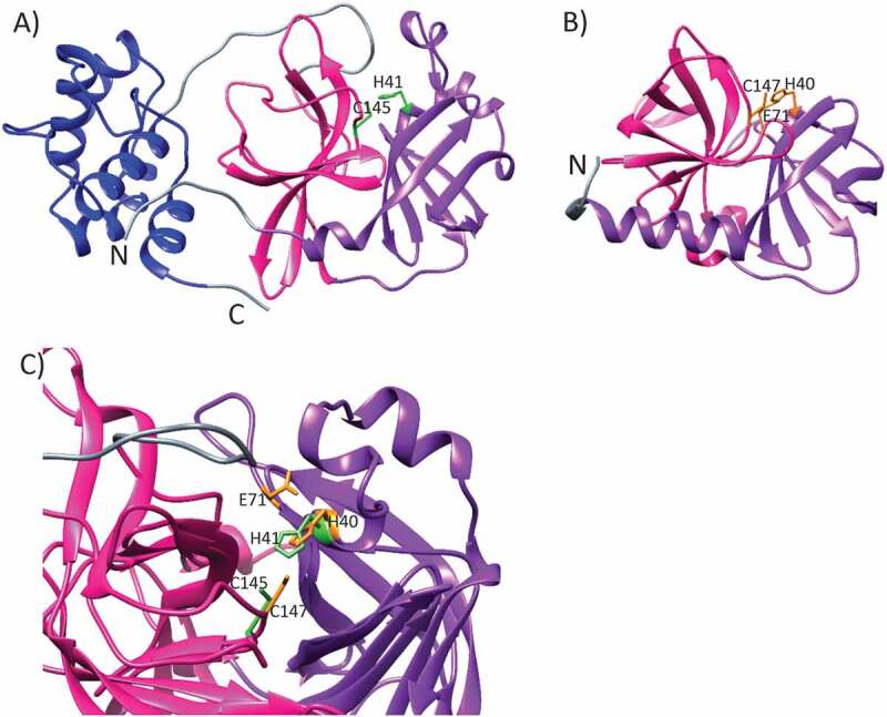
Figure 2.

The ribbon representations of (a) SARS-CoV-2 main protease (PDB id 7K3T, DOI: 10.2210/pdb7K3T/pdb) and (b) Human Enterovirus 71 3 C protease (PDB id 3OSY [43],). The two β-barrel domains I and II, participating in the catalytic activity, are shown with purple and pink, respectively. The dimerization domain, found in SARS-CoV-2 main protease only, is colored with blue. The linker regions are colored with gray. The side chains of residues critical for the catalytic activity are marked with light green and orange for SARS-CoV-2 Mpro and enterovirus 71 3 C protease, respectively. (c) SARS-CoV-2 Mpro superimposed with the enterovirus 71 3 C protease. The catalytic sites are denoted as in A) and B). It should be noted that for the sake of clarity, only one protomer of SARS-CoV-2 dimer is shown in A) and C)
