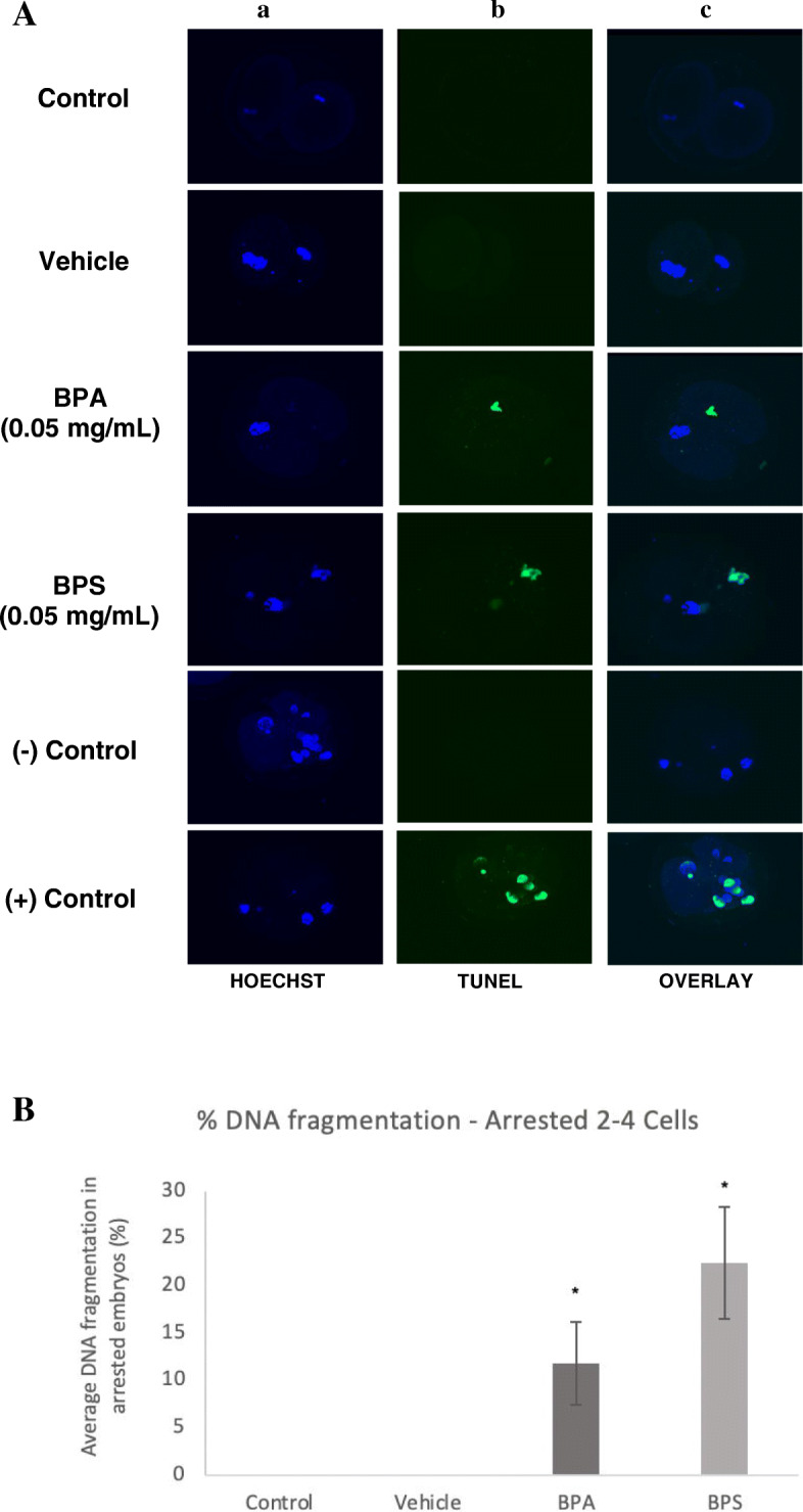
Fig. 4.

TUNEL assay of DNA fragmentation in arrested 2–4 cells. A Column a) is the nuclear counterstain (Hoechst), column b) is the FITC-labelled DNA fragmentation, column c) is an overlay of both. The last two rows display the negative and positive controls of the TUNEL assay. B Average DNA fragmentation in arrested 2–4 cells via TUNEL assay. Percentage of DNA fragmentation in each embryo determined as a ratio of the number of cells with DNA fragmentation to the total number of cells in the arrested embryos. * represents statistical significance p < 0.05 and error bars represent +/− SEM (n = 15 embryos per group)
