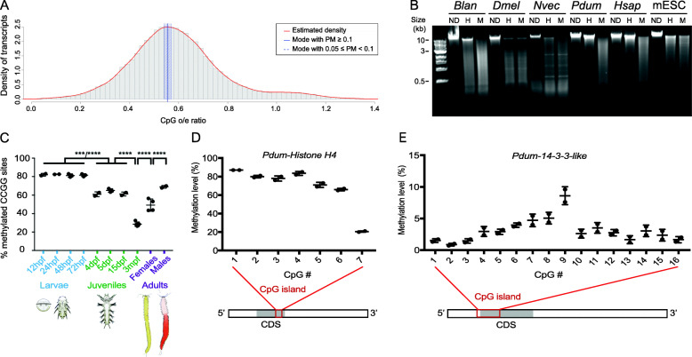
Fig. 1.
High-level and gene body CpG methylation in P. dumerilii. a Histogram of CpG o/e ratios of P. dumerilii transcripts. The red line indicates the estimated density, the vertical blue bar shows estimated mean value, and the shaded blue bar represents bootstrap confidence intervals of 95%. PM = probability mass. b Electrophoresis of non-digested (ND) genomic DNA (gDNA) or digested with HpaII (H) or MspI (M) from six different animal species with different methylation types. Sizes of fragments, in kilobase pairs (kb), are indicated to the left. Abbreviations: Blan = Branchiostoma lanceolatum; Dmel = Drosophila melanogaster; Nvec = Nematostella vectensis; Pdum = Platynereis dumerilii; Hsap = Homo sapiens; mESC = Mus musculus naïve embryonic stem cells. c Graphic representation of DNA methylation measured by LUMA at ten different stages of P. dumerilii life cycle (four larval stages, four juvenile stages, and adults (male and females); at least two biological replicates per stage and at least two technical replicates per biological replicate). Mean ± SD. One-way ANOVA, Tukey post hoc test (****: p < 0.0001, ***: p < 0.001). The raw data and results of all statistical tests can be found in Additional file 2: Table S2. hpf = hours post-fertilization; dpf = days post-fertilization; mpf = months post-fertilization. Drawings of larvae, juveniles, and adult worms are adapted from [35]. d, f Graphic representation of methylation levels of stretches of CpGs (CpG island) in two P. dumerilii genes, Pdum-Histone H4 (d) and Pdum-14-3-3-like (e), as defined by bisulfite pyrosequencing on DNA extracted from 72hpf larvae (two biological replicates and two technical replicates per biological replicate). Mean ± SD of two biological replicates is shown. A schematic representation of the localization of the studied CpG islands in the transcribed region of the two genes is also shown. CDS = coding sequence. Data shown in the graph and methylation levels at other developmental stages can be found in Additional file 2: Table S3

