Fig. 1.
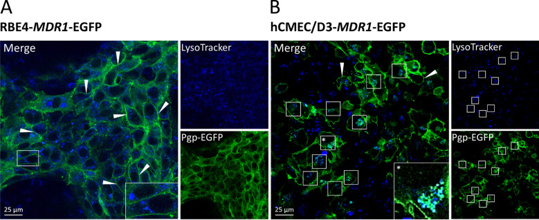
Subcellular localization of Pgp in RBE4- and hCMEC/D3-MDR1-EGFP cultures and endolysosomal staining. MDR1-EGFP transduced RBE4 (A) and hCMEC/D3 (B) cells grown on glass coverslips were treated with the fluorescent acidotropic LysoTracker probe to stain endolysosomal vesicles. Pgp localization visualized by green fluorescence of the EGFP protein tag and colocalization with endolysosomal vesicles was assessed by live-cell imaging and confocal laser scanning microscopy. Imaging revealed that, as expected, Pgp-EGFP (green) was localized at the cell surface in both cell lines (arrows). In hCMEC/D3-MDR1-EGFP cells, Pgp-EGFP (green) colocalized with LysoTracker (blue) stained intracellular vesicles (boxes in B), whereas this was hardly observed in RBE4-MDR1-EGFP cells. Boxes in the bottom right corner of the merged images show magnifications of Pgp subcellular localization in the respective cell line

