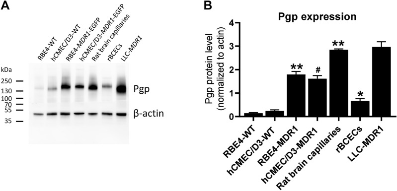
Fig. 3.
P-glycoprotein expression in hCMEC/D3 and RBE4 wildtype and MDR1-EGFP transduced cells in comparison to freshly prepared rat brain capillaries, primary cultured rat brain capillary endothelial cells, and MDR1-transfected LLC cells. Representative western blots (12 µg protein/sample) (A) and quantification (B) of Pgp expression. Pgp content of the immortalized BCEC lines (RBE4, hCMEC/D3) was determined in the presence of doxycycline. Data are presented as means + SEM; sample size (biological replicates) was 8 (RBE4-WT), 10 (hCMEC/D3-WT), 21 (RBE4-MDR1), 25 (hCMEC/D3-MDR1), 2 (rat brain capillaries), 6 (primary rBCECs), and 5 (LLC-MDR1), respectively. Data were analyzed by one-way ANOVA followed by Dunnett’s multiple comparisons test. Asterisks indicate significant differences of RBE4-MDR1, rat brain capillaries, and rBCECs to RBE4-WT (*P < 0.05; **P < 0.0001), whereas the hash sign indicated a significant difference between hCMEC/D3-MDR1 vs. hCMEC/D3-WT (#P < 0.0001)

