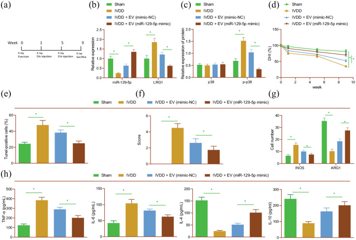
Figure 6.
Delivery of miR-129-5p by BMSC-derived EVs blunts the M1 polarization of macrophages and retards the progression of IDD in vivo. Rats were treated with EVs isolated from BMSCs that had been transfected with miR-129-5p mimic. (a) Flow chart for in vivo experiment. (b) miR-217 expression and LRG1 mRNA expression detected by RT-qPCR in rat NP tissues. (c) p38 phosphorylation level determined by Western blot analysis in rat NP tissues. (d) DHI in rat NP tissues. (e) TUNEL staining of NP cell apoptosis in rat IVD tissues. (f) Safranin-O/fast green staining of ECM degradation in rat IVD tissues. (g) Immunofluorescence staining of M1 marker iNOS and M2 marker ARG1 in rat IVD tissues. (h) Expression of TNF-α, IL-6, IL-4, and IL-10 detected by ELISA in rat serum. n = 8 for rats upon each treatment. *p < 0.05. The experiment was run in triplicate independently.

