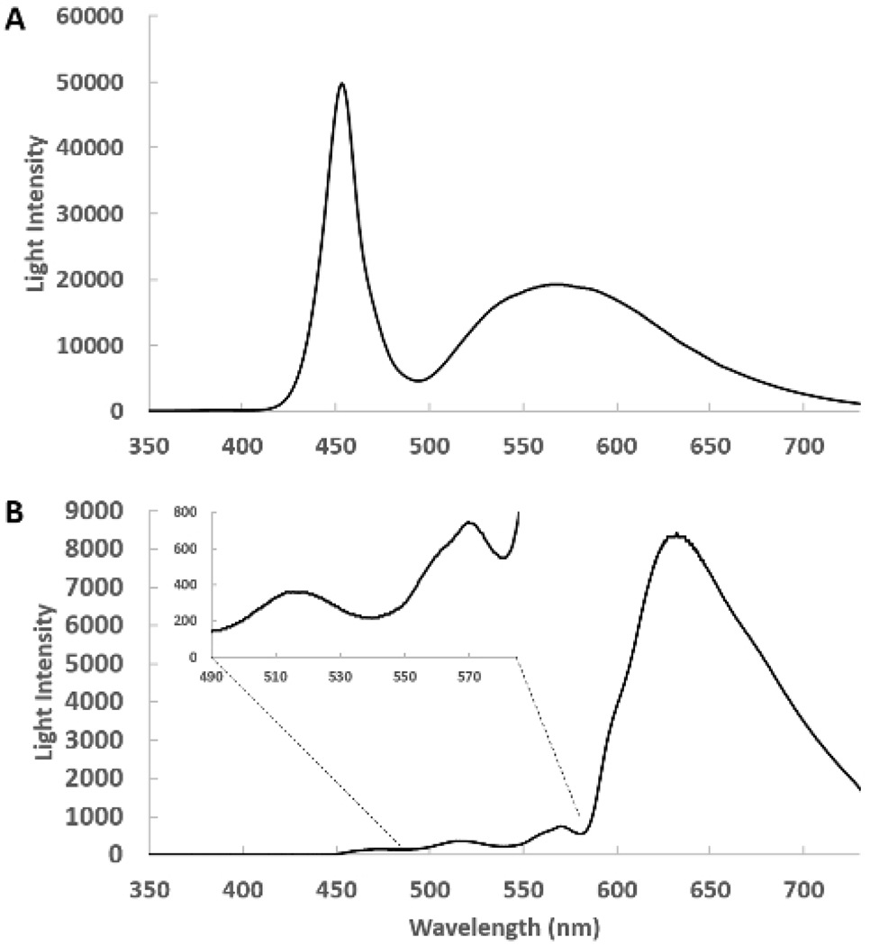
Figure 1: Spectra of the side-firing optical catheter.

(A) This is a spectrum of the emitted light from the remote light source through the catheter detected with the pickup fiber at about 1 cm from the catheter. In this geometry, the heart is absent and the intensity of the light source is tuned so that the detector does not saturate. (B) The side-firing catheter is inserted in the left ventricle and the transmitted light from the heart is collected and shown. The insert shows the 400 to 580 nm region expanded, revealing the complex transmission of light from this region. Please click here to view a larger version of this figure.
