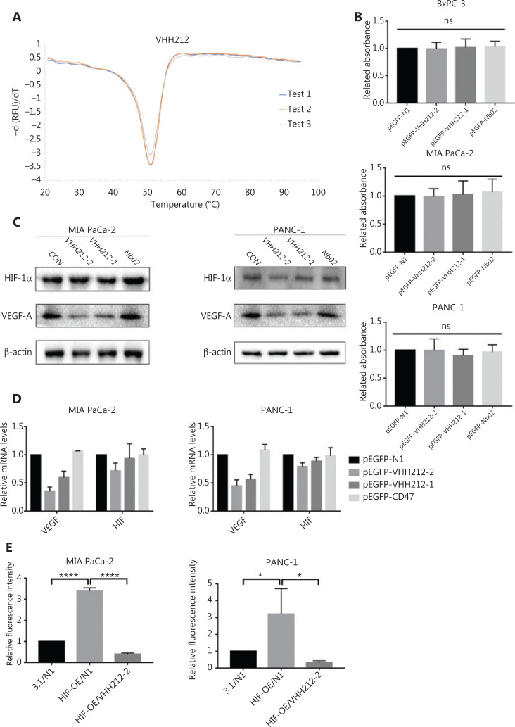
Figure 3.
VHH212 shows excellent thermal stability, low cytotoxicity, and the capacity to competitive inhibit the HIF-1 pathway. (A) An experiment to measure the thermal stability of VHH212. The Tm values were determined by melting curves using qPCR, and the value was average from 3 independent experiments. (B) Intrabodies do not have direct cytotoxic properties in vitro. The CCK8 assay was used to determine cell viability, and did not show a statistical difference between intrabodies and the vector control. (C) Western blot analysis of pancreatic cancer cell lines after hypoxic treatment. MIA PaCa-2 and PANC-1 cells were transfected with intrabodies and incubated under hypoxia (36 h), and total protein extracts were processed for Western blot using anti-HIF-1α, anti-VEGF-A, and anti-β-actin antibodies. (D) Quantitative real-time RT-PCR of pancreatic cancer cell lines after hypoxic treatment. MIA PaCa-2 cells were transfected, treated with hypoxia, and lysed in TRIzol for RNA extraction and analyzed for the mRNA expression of HIF-1α and VEGF by quantitative real-time RT-PCR. Data are expressed as the mean ± SEM. Columns, the mean of three experimental determinations; bars, standard deviation. (E) Luciferase analysis of MIA PaCa-2 and PANC-1 cells. The cells were transfected as described above. Relative luciferase analysis used the Dual-Luciferase Reporter Assay System, and the Renila vector was transfected as an internal control. Results are expressed as fold induction relative to cells transfected with the control vector (pcDNA3.1) after normalization to Renila activity. Columns, mean of 3 independent experiments; bars, standard deviation. (*P < 0.05) vs. the control. 3.1, pcDNA-3.1; N1, pEGFP-N1; VHH212, pEGFP-VHH212-2; HIF-OE, pcDNA-HIF-1α-OE. ****P < 0.0001.

