Figure 1.
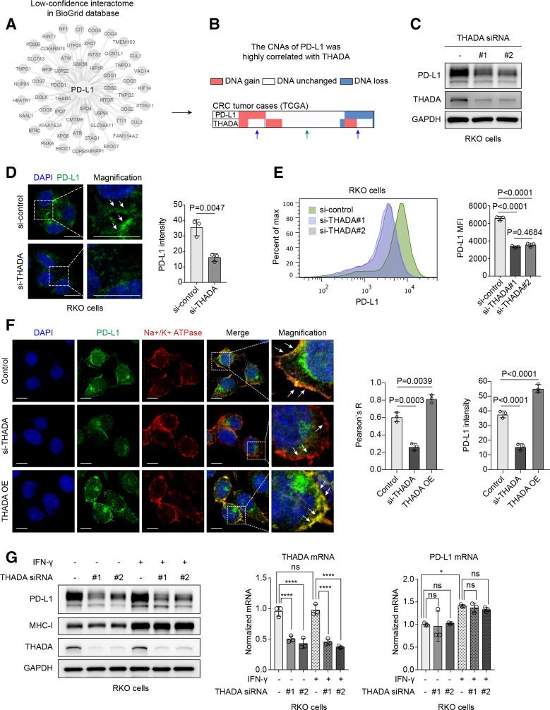
Thyroid adenoma associated gene (THADA) positively regulated programmed death-ligand 1 (PD-L1). Identification of THADA as a putative regulator of PD-L1. Combined analysis of a set of low-confidence interactors of PD-L1 summarized in the BioGrid database (A) with their gene copy number alterations (CNAs) in the colorectal cancer (CRC) data set of TCGA. The CNAs of THADA and PD-L1 were associated in the TCGA CRC data set (B). In CRC cases with no alteration in copy number of PD-L1 gene, THADA gene also tended to keep unchanged (green arrow), while in tumors with gained or loss of PD-L1 gene, THADA gene was prone to be amplified or diminished as well (blue arrow). (C) Western blot analysis showing the effect of THADA depletion by two distinct small interfering RNAs (siRNAs) on PD-L1 expression in RKO cells. The experiments were repeated three times independently with similar results. (D) Left, immunofluorescence assay showing RKO cells transfected with THADA siRNAs. Green, PD-L1; blue, DAPI nucleus staining. Scale bars, 10 µm. White dashed boxes indicate the representative fields to be magnified. Right, quantification of fluorescent intensity of PD-L1. Values are means±SD from n=3 independent experiments. Statistical differences were evaluated by two-sided Student’s t-test. (E) Left, flow cytometry showing the effect of THADA knockdown by two distinct siRNAs on PD-L1. Right, quantification of mean fluorescence intensity (MFI) of PD-L1. Values are means±SD from n=3 independent experiments. Statistical differences were evaluated by analysis of variance (ANOVA) post hoc test (Tukey). (F) Immunofluorescence assay showing PD-L1 localization on the plasma membrane in THADA depleted or overexpressed RKO cells. Scale bars, 10 µm. White dashed boxes indicate the representative fields to be magnified. Values are means±SD from n=3 independent experiments. Statistical differences were evaluated by ANOVA post hoc test (Tukey). (G) RKO cells transfected with two distinct THADA siRNAs and co-incubated with interferon (IFN)-γ (100 ng/mL, 24 hours), subjected to western blot analysis (left) and RT-PCR (right). Values are means±SD from n=3 independent experiments. Statistical differences were evaluated by ANOVA post hoc test (Tukey). *P<0.033; ****p<0.0001; ns, no significance.

