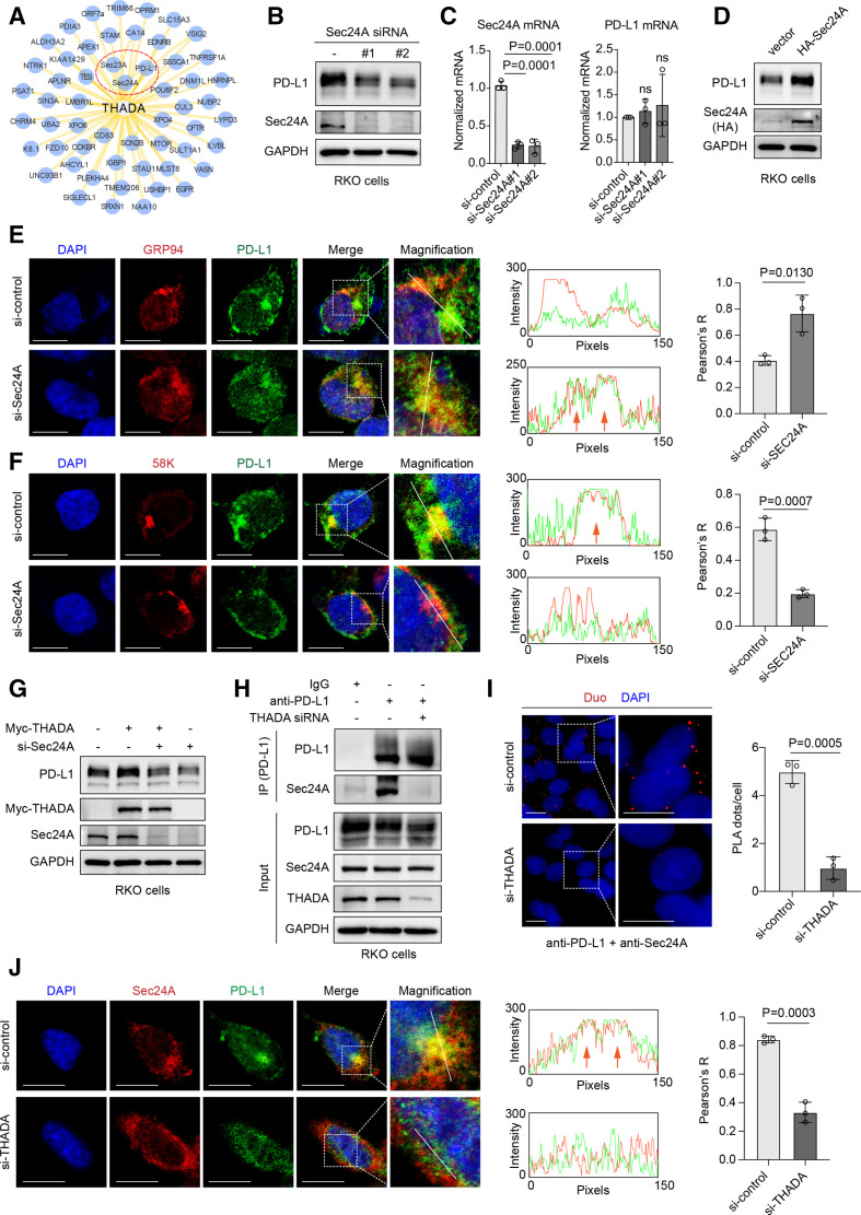
Figure 3.
Thyroid adenoma associated gene (THADA) required for Sec24A-dependent vesicle trafficking of programmed death-ligand 1 (PD-L1). (A) A set of low-confidence interactors of THADA summarized in the BioGrid database. RKO cells transfected with two distinct Sec24A small interfering RNAs (siRNAs) and subjected to western blot analysis (B) and RT-PCR (C), respectively. Values are means±SD from n=3 independent experiments. Statistical differences were evaluated by analysis of variance (ANOVA) post hoc test (Tukey); ns, no significance. The experiments were repeated three times independently with similar results. (D) Western blot analyses showing the effect of Sec24A overexpression (HA-Sec24A) on PD-L1 in RKO cells. The experiment was repeated three times independently with similar results. Left, immunofluorescence assays showing the co-localization between PD-L1 and GRP94 (E)/58K (F) in RKO cells transfected with Sec24A siRNAs. Scale bars, 10 µm. White dashed boxes denote the representative fields to be magnified. Middle, the intensity profiles of PD-L1 and GRP94 (E)/58K (F) along the white line. Orange arrows denote the co-localization. Right, values are means±SD from n=3 independent experiments. The p values were evaluated by two-sided Student’s t-test. (G) RKO cells transfected Myc-tagged THADA and Sec24A siRNAs as indicated and subjected to western blot analysis with indicated antibodies. (H) Co-immunoprecipitation (Co-IP) assay showing the interaction between PD-L1 and Sec24A in RKO cells transfected with THADA siRNAs. The experiments were repeated three times independently with similar results. (I) Duolink assay showing the interaction of endogenous PD-L1 and Sec24A proteins in RKO cells. Red dots indicating the binding of two proteins. Nuclei were stained with DAPI. Scale bar, 10 µm. Values are means±SD from three random fields in each treatment. Statistical differences were evaluated by two-sided Student’s t-test. (J) Left, immunofluorescence assay showing the co-localization between PD-L1 and Sec24A in RKO cells transfected with THADA siRNAs. Scale bars, 10 µm. White dashed boxes denote the representative fields to be magnified. Middle, the intensity profiles of PD-L1 and Sec24A along the white line. Orange arrows denote the co-localization. Right, values are means±SD from n=3 independent experiments. The p values were evaluated by two-sided Student’s t-test.

