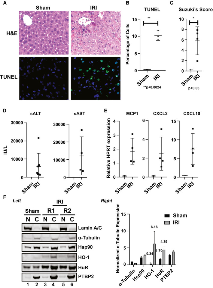
Figure 1.

HO‐1 and HuR protein levels are up‐regulated in a mouse warm hepatic IRI model. Livers in C57/BL mice were subjected to 60 minutes of portal triad blockage and subsequent reperfusion for 6 hours. (A) Representative hematoxylin and eosin (H&E) and (B) terminal deoxynucleotidyl transferase–mediated deoxyuridine triphosphate nick‐end labeling (TUNEL) staining (n = 3; original magnification, ×100; **P < 0.0024). (C) Suzuki's score (*P < 0.05; IRI vs. Sham) of the liver sections, as well as (D) sALT/sAST levels (n = 4–7/group; mean ± standard error, IU/L) and (E) mRNA levels of chemokines MCP1, CXCL2, and CXCL10 are shown. (F) Left: representative fractionation of nuclear and cytoplasmic proteins from liver tissues obtained from animals exposed to warm IRI. Lamin A/C (nuclear marker), α‐tubulin (cytosolic marker), Hsp90 (stress‐response comparison control), HO‐1 (subject of study), HuR (subject of study), and PTBP2 (downstream effector of HuR) are shown. R1 and R2 are replicates 1 and 2. Right: unpaired two‐tailed Student t test of representative samples presented were calculated relative to α‐Tubulin cytoplasmic expression. Data shown are mean ± SEM; n = 2 (repeated at least three independent times). HPRT, hypoxanthine‐guanine phosphoribosyltransferase.
