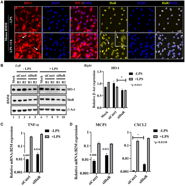
Figure 4.

Silencing of HuR enhances proinflammatory signatures in BMMs. (A) Mouse (C57BL/6) BMM was subjected to LPS endotoxin for 3 hours followed by HO‐1 or HuR immunofluorescence staining. White arrows signify areas of cytoplasmic distribution. (B) Left: total lysates from LPS‐conditioned BMM were treated with siControl (siCntrl) or siHuR RNAs and probed by western blot for differences between HO‐1, HuR, and β‐Act as a loading control. Samples separated by a border were analyzed on the same western blot. Equal protein amounts (25 μg) from protein lysates were loaded on each lane. Right: quantitation of HO‐1, data shown are mean ± SEM, *P < 0.0333; siHuR +LPS versus siCntrl +LPS. (C) Inflammatory cytokine analyses of TNF‐α by qPCR. (D) mRNA coding for inflammatory chemokines (MCP1, CXCL2). Data were normalized to B2M gene expression, n = 3. Act, actin; DAPI, 4′,6‐diamidino‐2‐phenylindole.
