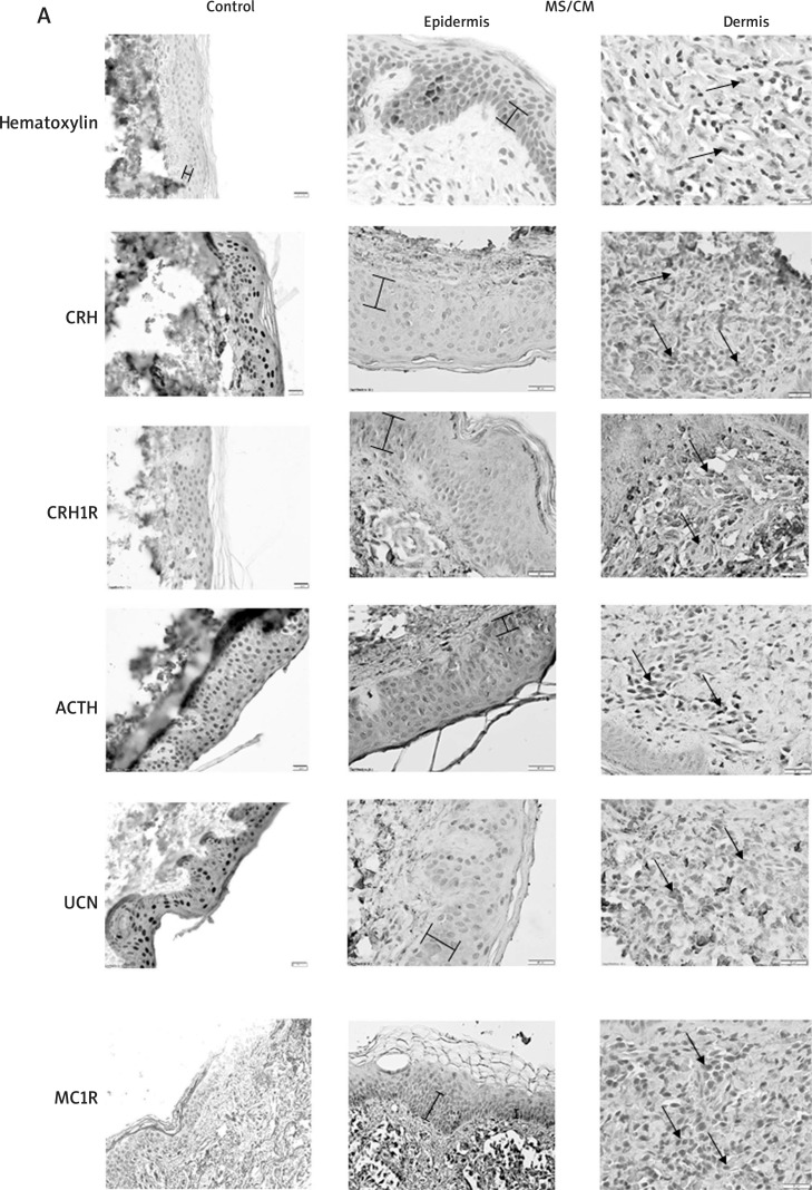
Figure 3 A.
Immunodetection of selected neuropeptides and their receptors in positive IHC labelling is represented in brown. The left panel represents healthy controls, middle – representative epidermis of SM/CM patients, right – representative dermis of SM/CM patients. Braces mark the range of pigment; arrows indicate positive NMC cells. Haematoxylin stained samples, where primary antibodies omitted, serve as negative controls. See Tables 3 or 4 for details.

