
Figure 2 .
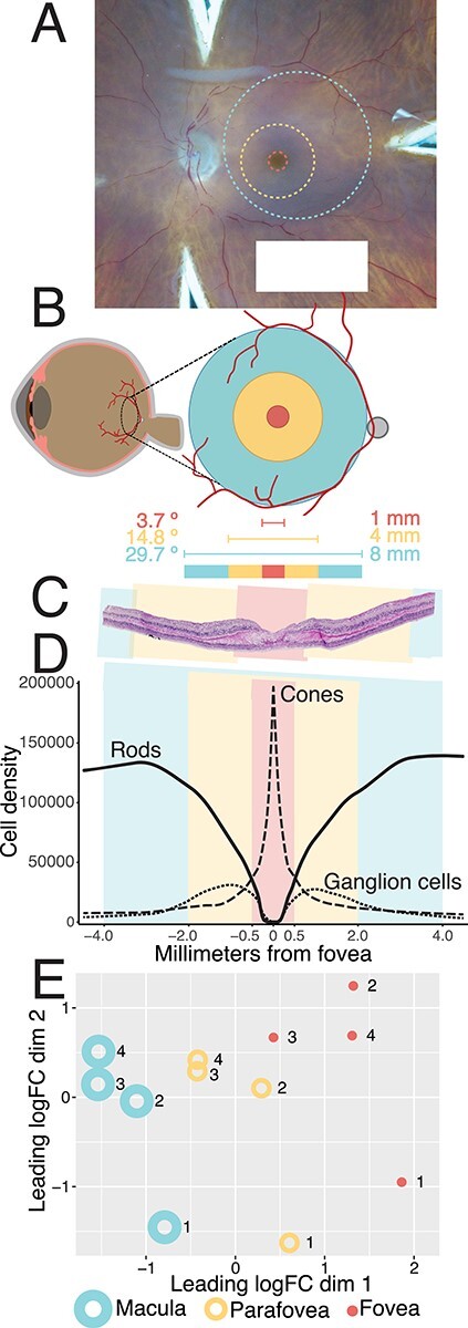
Comparing gene expression in foveal (1 mm), parafoveal (4 mm) and macular (8 mm) retina. (A) A hypothetical 1 mm foveal-centered punch (red), 4 mm parafoveal punch (yellow), and 8 mm macular punch (blue) are overlayed on a gross photo of a human donor eye. Punches were acquired concentrically; however, the position of the 8 mm macular punch was slightly adjusted on a case-by-case basis to avoid the optic nerve head. (B) The contribution of each punch to the visual field was estimated (12). (C) Hematoxylin and eosin histological staining of fovea-centered retina. A perfectly centered 1 mm foveal punch (red) includes the entire foveal pit. (D) Density of cones, rods and RGCs across each of the investigated regions of macular retina (14). Cones reach their peak density within the foveola, which is completely captured in a perfectly centered 1 mm punch. (E) After bulk RNA-sequencing of four human donors, multidimensional scaling reveals separation of macular, parafoveal, and foveal retinal samples, suggesting that each region has a unique transcriptome.