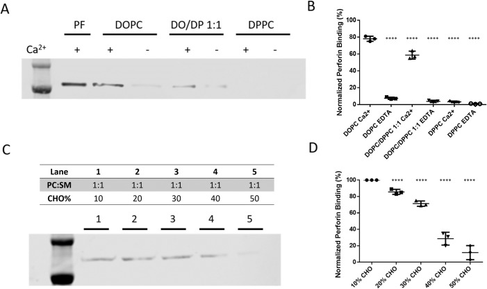
Fig 5. Perforin membrane binding is prevented by the presence of densely packed lipid.
Differently packed liposomes were created by either pure or a ratio of DOPC and DPPC, which were coincubated with perforin. Levels of membrane-bound perforin were evaluated by western blot analysis (A, representative of 3 independent repeats). Since membrane binding of perforin requires the presence of Ca++, this was either added at 20 mM concentration or depleted as specified in the blot and quantification. As a control, pure perforin (PF) was evaluated without liposomes. Three independent experiments were performed, and the quantitative values for each via densitometry were plotted with mean ± SD shown (B). Using Student t test, the mean values were different from DOPC Ca++ p < 0.0001, **** (C) Differently packed PC:SM:CHO liposomes were created with 1:1 PC:SM and increasing percentages of CHO (as specified above each lane) and coincubated with perforin in the presence of Ca++. Levels of membrane-bound perforin were evaluated by western blot analysis using anti-perforin antibody (clone D48) (representative result shown). Three independent experiments were performed, and the quantitative values for each via densitometry were plotted with mean ± SD shown (D). Using Student t test, the mean values were different from 10% CHO p < 0.0001, ****. CHO, cholesterol; DOPC, dioleoyl phosphatidylcholine; DPPC, dipalmitoyl phosphatidylcholine; PC, phosphatidylcholine; SM, sphingomyelin.

