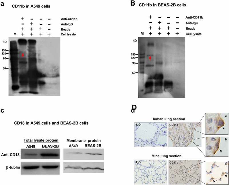
Figure 1.

Expression of CR3 (CD11b/CD18) in airway epithelial cells. Total cell lysates of either A549 cells (a) or BEAS-2B cells (b) were incubated with protein A/G agarose beads and CD11b expression were analyzed by immunoprecipitation with anti-CD11b mAb (ab52478). The red arrows in A and B indicated the band of CD11b protein. (c) The total lysate protein and membrane protein of A549 cells and BEAS-2B cells were also analyzed for CD18 expression by immunoblotting using an anti-CD18 antibody (ab52920). β-tublin was used as a loading control. (d) Representative paraffin sections of human lung tissues from surgical specimens from seven lung cancer patients and mice lung tissues from normal C57BL/6 wild-type mice were stained for CD11b using anti-CD11b mAb (ab52478) and photographed at an optical magnification of 100. The arrows (black) indicated the staining of CD11b on lung epithelial cells. In human lung section, scale bar = 9.95 μm (IgG and CD11b). In mice lung section, scale bar = 50 μm (IgG and CD11b). a, b and c were the amplification of the specified area respectively. Images of immunohistochemical staining and immunoblots shown here are characteristic of 3 independent experiments
