
Figure 3.
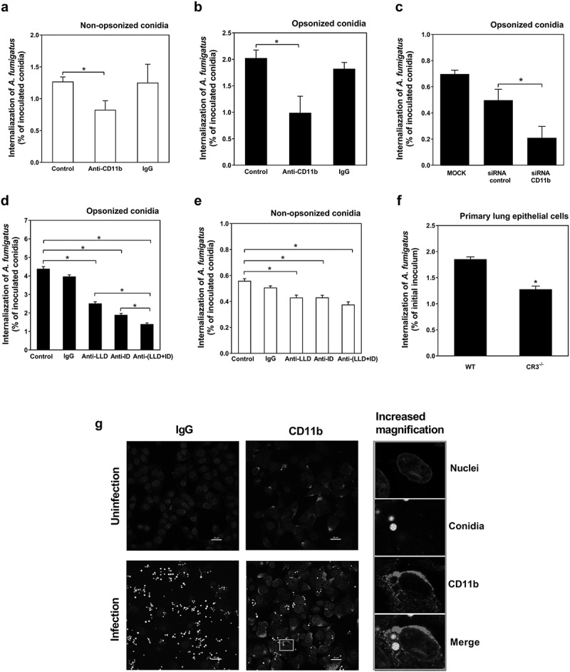
CD11b are involved in the internalization of A. fumigatus conidia into lung epithelial cells by opsonization. (a-f) The internalization of A. fumigatus ATCC13073 conidia into A549 cells was analyzed by nystatin protection assay, MOI = 10, incubation time = 6 h. A549 cells were pre-treated with or without anti-CD11b monoclonal antibody (NBP1-28,423, 10 μg/ml) and isotype control IgG for 30 min at 37°C, respectively, then infected with the resting conidia of A. fumigatus ATCC13073 which were opsonized without (a) or with (b) human serum. (c) A549 cells were transfected siRNA for negative control and CD11b by lipofectamin 2000 for 48 h, respectively, then infected with the resting conidia of A. fumigatus ATCC13073 which were opsonized with human serum. A549 cells were pre-treated with or without anti-CD11b either I-domain (anti-ID, 10 μg/ml) and/or lectin-like domain (anti-LLD, 10 μg/ml) monoclonal antibody and isotype control IgG for 30 min at 37°C, respectively, then infected with the resting conidia of A. fumigatus ATCC13073 which were opsonized without (d) or with (e) human serum. (f) Primary pulmonary epithelial cells from wild type C57 mice and CR3−/- mice were stimulated with the resting conidia of A. fumigatus ATCC13073 for 6 h. Data were representative of 3–4 independent experiments or from means ± s.e.m of four independent experiment. The multiple t-test was performed, *p < 0.05. (g) The internalization of A. fumigatus into A549 cells and the expression of CD11b were monitored after staining by fluorescence microscopy using fluorescence microscope Olympus BX51 (green, A. fumigatus conidia; red, CD11b). A549 cells were infected with or without the resting conidia of A. fumigatus 13073 stably expressing green fluorescence protein (MOI = 10) for 6 h. Then the cells were stained with anti-CD11b primary antibody(ab75476) and Alexa Fluor 594 goat anti-rabbit IgG secondary antibody(red), DAPI (stain nuclei, blue). The images were processed with Image-Pro Express, Scale bar was shown on the images