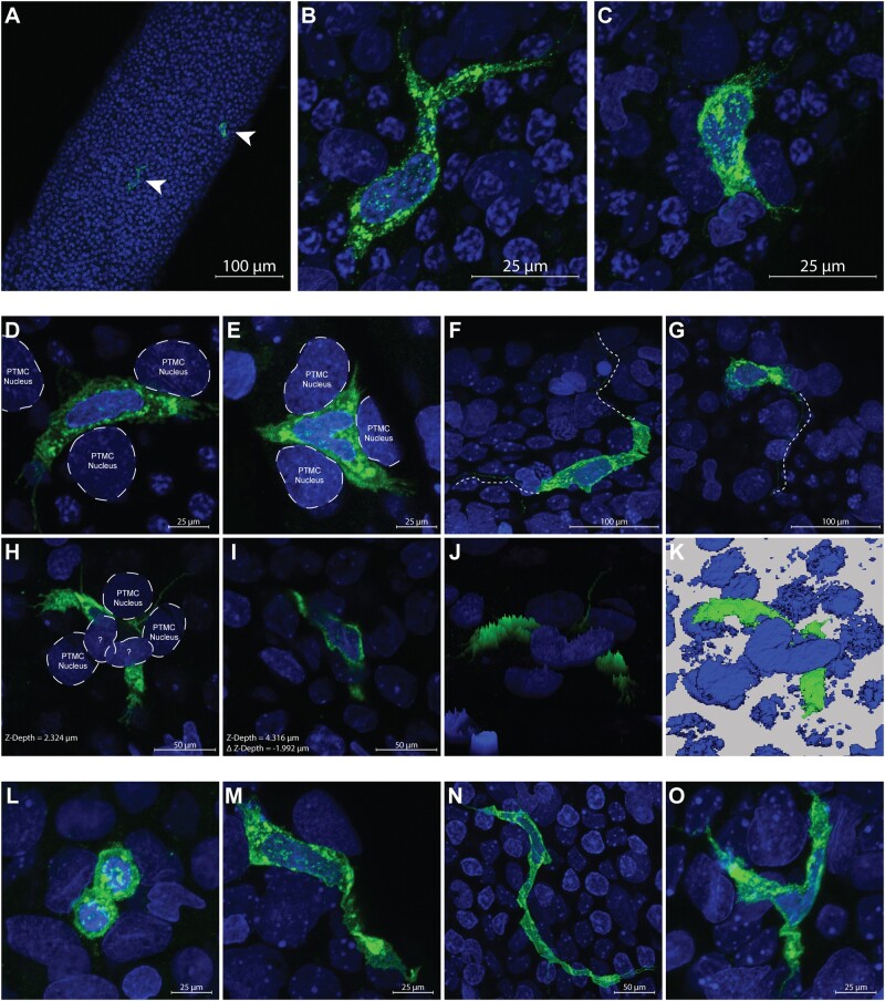
Figure 1.
A, major histocompatibility complex class II (MHCII+) peritubular macrophages vehicle control (VEH—green—arrow heads) are shown on the surface of a whole seminiferous tubule at 20×; scale bar = 100 μm. B and C, Up close of (A; 63×; scale bar = 25 μm) confocal images show 2 MHCII+ peritubular macrophages from VEH animals with heterogeneous morphology on the surface of a seminiferous tubule. D and E, Two separate macrophages D (from VEH) and E (from mono-(2-ethylhexyl) phthalate [MEHP]) are shown at high magnification (63×; scale bar = 25 μm) nestled between the nuclei of what appear to be peritubular myoid cells (PTMCs), identified by their location and unique nuclei morphology. The macrophage cell surface marker MHCII can be seen to occupy space in the same plane and between the presumed PTMC nuclei. F and G, Long (>100 μm) extensions can be seen protruding bidirectionally or unidirectionally (respectively) from MHCII+ cells from VEH animals (offset dashed/dotted line), sometimes meandering under the nuclei of other cells (63×; scale bar = 25 μm). H and I, An MHCII+ cell from MEHP group is shown in 2 different scanning planes (H = Z-Depth 2.324 μm and I = Z-Depth 4.316 μm) that is nestled between 3 presumed PTMC nuclei and under the nuclei of 2 cells unknown type (63×; scale bar = 50 μm). J, A 3D brightness histogram and K, a reconstructed 3D image shows an alternative view of an MHCII+ cell under the nuclei of 2 cells of unknown type from MEHP-treated animals. The various morphologies of MHCII+ peritubular macrophages observed on the surface of seminiferous tubules is shown; (L) circular from VEH group (63×; scale bar = 25 μm); (M) spindeloid from MEHP group (63×; scale bar = 25 μm); (N) elongated from MEHP group (63×; scale bar = 50 μm); (O) stellate from MEHP group (63×; scale bar = 25 μm).

