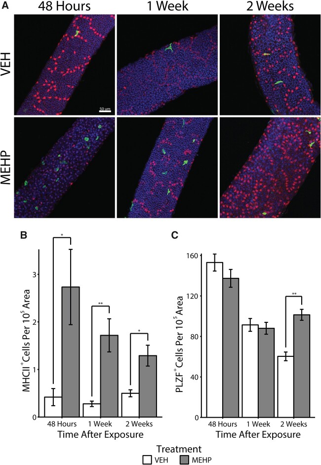
Figure 3.
A, Representative micrographs of major histocompatibility complex class II (MHCII+; green) peritubular macrophages are shown on the surface of seminiferous tubules in close proximity to PLZF+ (red) spermatogonia in both vehicle control (VEH) and mono-(2-ethylhexyl) phthalate (MEHP)-treated rats after 48 h after exposure to MEHP. All images in this figure were taken at 20×. The scale bar (50 μm) represented in the top left panel applies to all images in this figure. B, The number of MHCII+ cells, normalized to tubule area, is shown. *p < .01 and **p < .001. C, The number of PLZF+ spermatogonia in VEH and MEHP-treated male rats is shown normalized to tubule area (**p < .001).

