Figure 1.

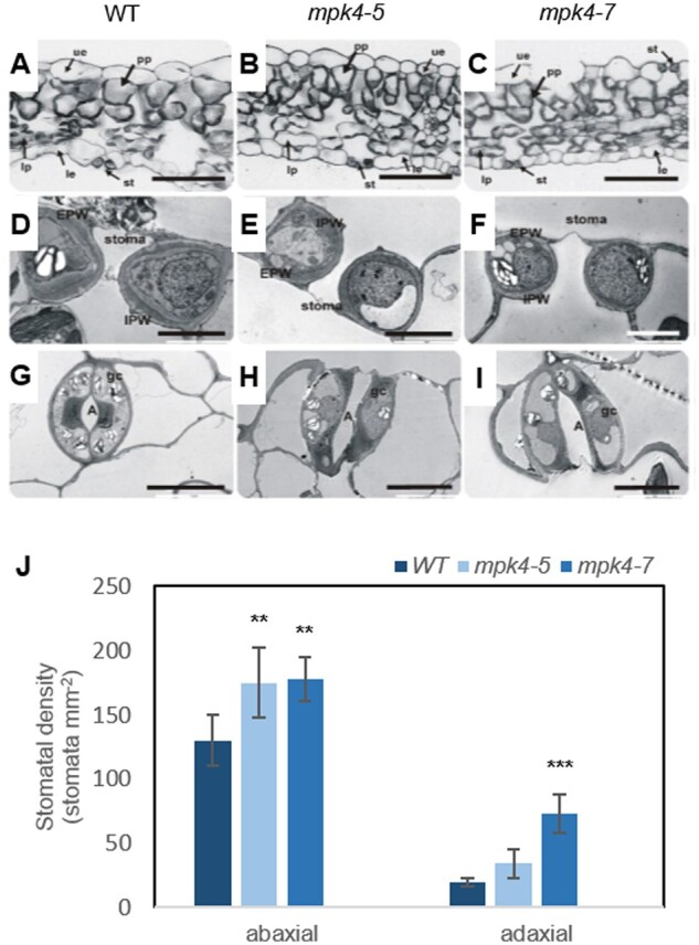
Leaf development, stomatal ultrastructure, and stomatal density were dependent on MPK4. Microscopic analysis was performed for WT and transgenic mpk4-5 and mpk4-7 plants. A–C, Light micrographs of a leaf cross-section. le, lower epidermis; lp, lacunous parenchyma; st, stoma; ue, upper epidermis. D–F, Transmission electron micrographs showing a median transverse section of a stoma. G–I, Paradermal sections of guard cells from the le. A, aperture; EPW, external periclinal wall; IPW, internal periclinal wall. Scale bars: (A–C) 50 μm; (D–F) 5 μm; (G–I) 10 μm. J, Mean stomatal density ± sd (n = 3) of the epidermal layer in adaxial (upper) and abaxial (lower) leaf surfaces. The asterisks indicate significant differences from the WT revealed by Tukey’s HSD test; **P < 0.01, ***P < 0.001.
