Figure 2.
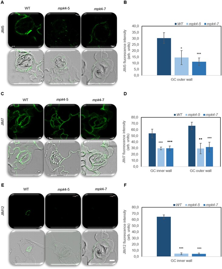
Pectin composition and extensin level in WT and transgenic mpk4-5 and mpk4-7 GC walls. The green signal is the signal from the antibody conjugated to FITC. An overlay between the green antibody signal and bright-field is also presented. The JIM5 antibody (A–B) recognizes the HG domain of pectic polysaccharides, partially methyl-esterified epitopes of HG, and unesterified HG. The JIM7 antibody (C–D) recognizes the HG domain of pectic polysaccharides, partially methyl-esterified epitopes of HG but not unesterified HG. The extensins are indicated by the JIM12 antibody (E–F). Scale is uniform for each photo, scale bar: 2 μm. Control without primary antibody was made for each type of sample to confirm the lack of signal (Supplemental Figure S3). The presented pictures are representative of 10 analyses ±sds (n = 30). Fluorescence was measured using Leica confocal software and is expressed as arbitrary units. The asterisks indicate significant differences from the WT revealed by Tukey’s HSD test; *P < 0.05, **P < 0.01, ***P < 0.001.

