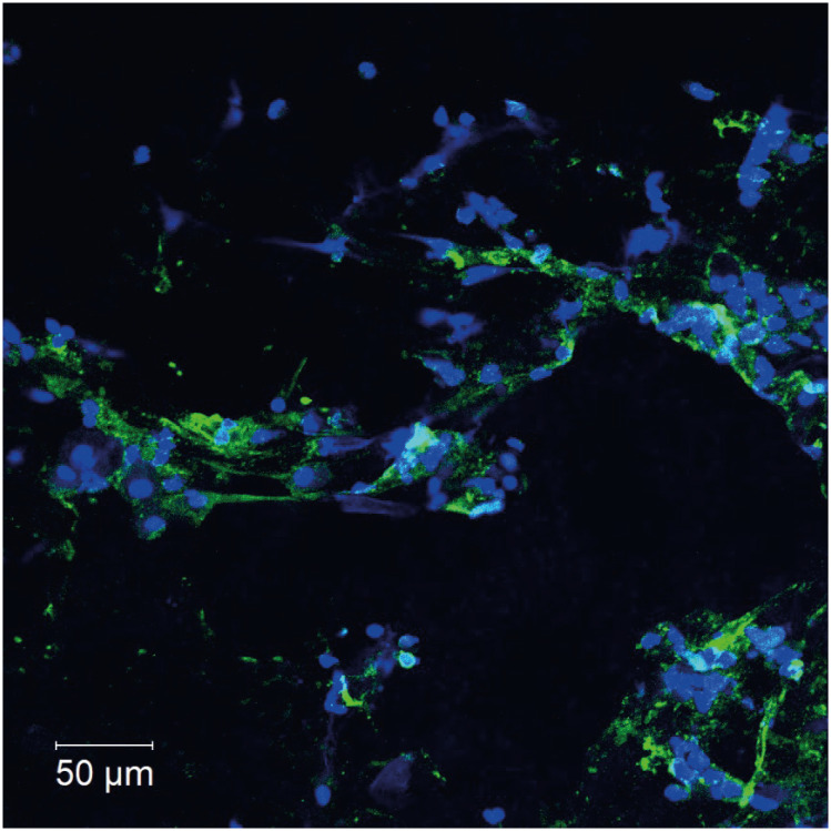
Fig. 3. Citrullinated histone H3 (CitH3) immunostaining to detect eosinophil extracellular traps in bronchoalveolar lavage fluid (BALF).
Immunofluorescent staining of citrullinated histone H3 (green) and DNA (blue) in BALF from this patient, which was collected before this patient received steroid treatment. The BALF was applied to slides, fixed with paraformaldehyde, incubated for 15 minutes in Tris-EDTA buffer in a microwave oven, blocked with 10% bovine serum albumin and 1% saponin coating phosphate-buffered saline for 30 minutes, and finally incubated with primary rabbit anti-CitH3 mAb (10 μg/mL, Abcam, Cambridge, UK), 90 minutes at room temperature [RT]) and with Alexa-488-conjugated antibodies (Life Technologies, Waltham, MA, USA; 30 minutes at RT) for the secondary incubation. Isotype-matched control antibodies and Hoechst 33342 (Life Technologies) were used for each experiment. Images were obtained using a LSM780 confocal microscope (Carl Zeiss, Oberkochen, Germany). CitH3-positive extracellular traps spread in a network, confirming that some cells have ETosis. Scale bars: 50 µm.

