
Figure 5.
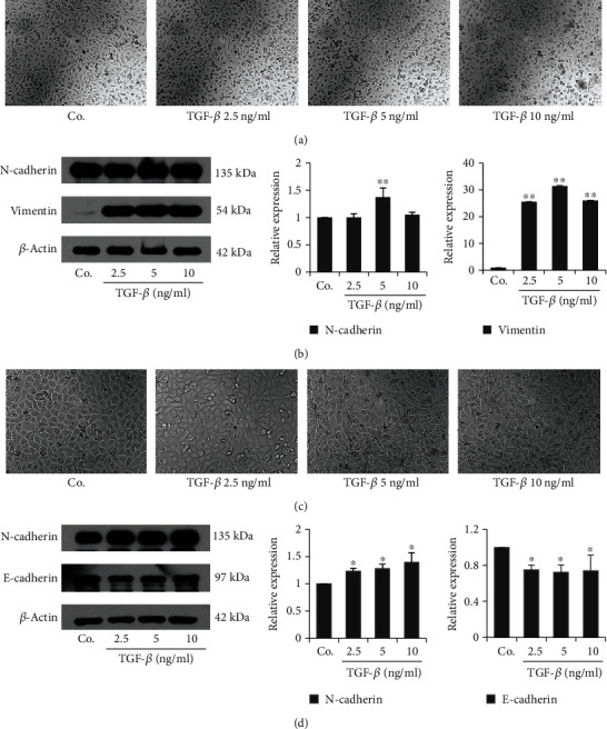
Establishment of TGF-β-induced EMT model of BC cells. T24 (a, b) and 5637 (c, d) were treated with TGF-β for 24 h. The morphological changes were observed under a phase-contrast microscope and photographed (100x). The level of EMT markers was examined by western blotting and the results were quantified. The data were presented as mean ± SD (n = 3). ∗P < 0.05, ∗∗P < 0.01 vs. control group.