Figure 3.

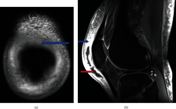
(a) MRI coronal view reveals an area of hypodensity in the prepatellar bursa indicating bursitis (blue arrow). (b) Sagittal T1 view reveals prepatellar bursitis (blue arrow) and abscess (red arrow).

(a) MRI coronal view reveals an area of hypodensity in the prepatellar bursa indicating bursitis (blue arrow). (b) Sagittal T1 view reveals prepatellar bursitis (blue arrow) and abscess (red arrow).