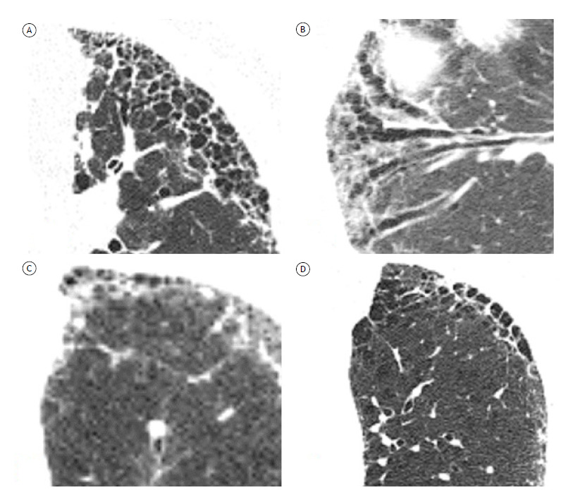
Figure 2. Axial HRCT scans with lung window settings, showing fibrosing interstitial lung disease in different patients. In A, typical honeycombing, presenting as multiple layers of cysts. In B, traction bronchiectasis in an oblique coronal plane. Note the usefulness of multiplanar reconstruction in differentiating between traction bronchiectasis and honeycombing. In C, early honeycombing, presenting as a single layer of cysts. In D, note the difficulty in differentiating between honeycombing and paraseptal emphysema.

