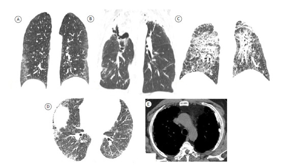
Figure 4. Axial HRCT scans of the chest showing features related to different fibrosing diseases. In A, features suggestive of nonspecific interstitial pneumonia, with a predominance of ground-glass opacities. In B, features of pleuroparenchymal fibroelastosis confirmed by histopathology showing predominantly apical fibrosing disease with upper lobe volume loss and upward hilar retraction. In C, scan of a patient with sarcoidosis, showing fibrosing disease with an upper-lobe predominance. In D and E, scans of a patient with fibrosing lung disease caused by exposure to asbestos. Note the presence of pleural plaques (arrows in E).

