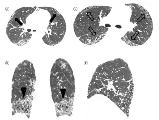
Figure 6. Specific CT signs of fibrosis in connective tissue disease-associated fibrosing interstitial lung disease. In A and B, the “anterior upper lobe” sign (arrows) in a patient with rheumatoid arthritis. In C, the “four corners” sign (open arrows)-fibrosis focally involving the bilateral anterolateral upper lobes and posterosuperior lower lobes-in a patient with systemic sclerosis. In D, the “straight-edge” sign (arrowheads)-isolation of fibrosis to the lung bases with sharp demarcation in the craniocaudal plane. In E, the “exuberant honeycombing” sign-extensive honeycombing constituting most of the fibrotic portions of lung.

