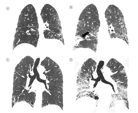
Figure 8. Coronal reconstructions of HRCT scans showing complications of fibrosing interstitial lung disease in two patients. In A and B, scans of a patient with progressive idiopathic pulmonary fibrosis. Initial CT scan (in A) and follow-up CT scan at seven years and 10 months (in B), showing disease progression and an irregular subpleural expansile neoplastic lesion in the right lower lobe, diagnosed as adenocarcinoma (arrow in B). In C and D, scans of a patient with progressive idiopathic pulmonary fibrosis. Initial CT scan (in C) and follow-up CT scan at 15 months (in D), showing disease progression and new bilateral ground-glass opacities, characterizing acute exacerbation of idiopathic pulmonary fibrosis.

